Bubble-based diagnosis of bacterial and viral diseases
-
- from Shaastra :: vol 03 issue 06 :: Jul 2024
Researchers discover an in situ diagnostics technique that uses microbubbles to detect bacterial and viral diseases.
Testing blood or urine samples can be a lengthy process. This delay can be critical for patients when time is of the essence. Researchers have discovered an in situ diagnostic technique that delivers immediate results by applying a method called microbubble lithography. The research, led by Ayan Banerjee from the Indian Institute of Science Education and Research (IISER) Kolkata, was published in the journal Small (bit.ly/microbubble-small).
Over a decade ago, Banerjee and his student Basudev Roy (now an Associate Professor at the Indian Institute of Technology Madras who was not involved in the study) had serendipitously discovered that microbubbles formed when a sharp laser was used on some materials. They also observed that moving the laser source caused the bubbles to leave a trail, a process they termed microbubble lithography.
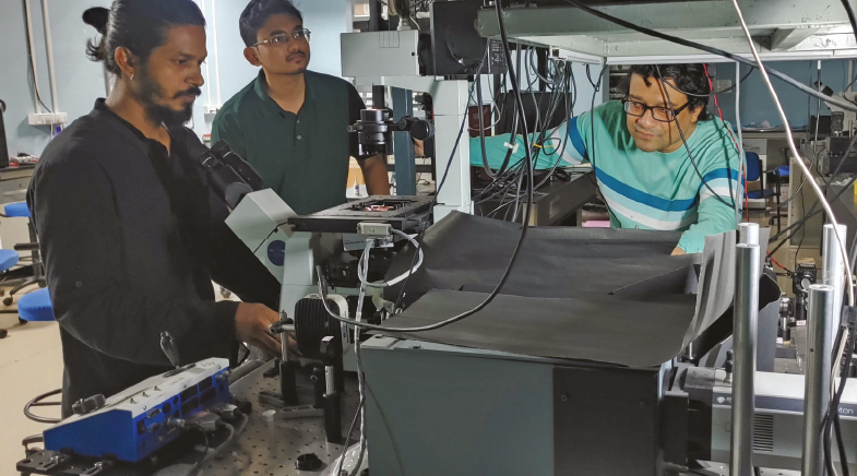
Now, Banerjee and his IISER team focused a high-intensity laser beam on a tiny sample of biomolecules, about a picolitre (one trillionth of a litre), placed on a coverslip. The laser locally boiled the water molecules contained in the sample, creating bubbles as small as one micron (one millionth of a meter). By moving the laser, the researchers created a path of bubbles, "similar to the trajectory made by jet planes," explains Banerjee.
The IISER team focused a high-intensity laser beam on a tiny sample of biomolecules.
The researchers then created parallel lines of antibody proteins for diagnostic tests to detect bacterial and viral diseases. When a patient's sample is added to the covered slits with these antibody patterns, a colour change occurs if the sample contains the target virus, like a pregnancy test. "I thought the real game-changer would be if we could pattern proteins," Banerjee remarked. This process will make it possible to detect multiple viruses and bacteria simultaneously. The researchers tested their method by detecting Escherichia coli bacteria that cause diarrhoea and type A influenza virus. But shining an intense laser on biomolecules like proteins could destroy the cells. So, Banerjee's group infused other, 'linker' molecules with biomolecules to help them withstand the heat generated by the laser.
The researchers aim to create affordable 3D-printed diagnostic test kits capable of detecting multiple pathogens, similar to the widely available Rapid Antigen Test. "It will be much faster and much more targeted," says Banerjee. The microbubble lithography technique has been used to pattern diverse mesoscopic materials for applications like plastic electronics, catalytic chips, and biosensing. However, the IISER group is the first to apply this technique to biomolecules.
"The work is very inspiring," said Sangram Bagh, a biophysicist at the Saha Institute of Nuclear Physics, Kolkata. "(It) has enormous potential applications, including developing new drug-screening platforms, detecting pathogens, and generating living materials," says Bagh.


On a hydrogen hunt
Project PRATUSH is proposed to fly to the far side of the Moon to observe signals from an adolescent universe.

Wonder water
Plasma-activated water can kill harmful microbes and aid in faster growth of crops.


Small, large and brainy
Ideas that power sectors, cool areas and relieve pain.

Have a
story idea?
Tell us.
Do you have a recent research paper or an idea for a science/technology-themed article that you'd like to tell us about?
GET IN TOUCH