Handheld device detective
-
- from Shaastra :: vol 04 issue 11 :: Dec 2025
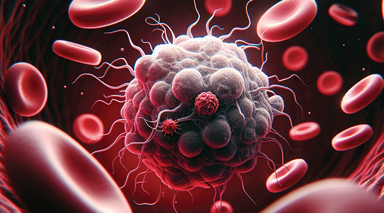
Researchers have devised a pen-like tool that works by shining light on tissues.
Researchers have developed a handheld tool for distinguishing malignant breast cancer cells from adjacent healthy cells. The pen-like device, with multiple near-infrared (NIR) light-emitting diodes (LEDs), identifies healthy and malignant cells based on their lipid and collagen content, reports a study in Scientific Reports (bit.ly/Cancer-Device).
Current breast cancer diagnoses and margin assessments — margin is the border of healthy tissue around the tumour — rely on lab-based histopathological studies. Researchers study a tissue to assess whether the cells are healthy or malignant. In tumour excision surgeries, the margins must be examined to ensure that the entire tumour has been excised. About 10-21% patients require a second surgery to achieve clear or negative margins, studies show. Second surgeries delay post-surgical treatments and have a greater chance of cancer recurrence and higher mortality risk.
The current device may help detect cancer cells in real time. It works by shining light at three wavelengths — 850 nm, 940 nm, and 1050 nm — chosen because they correspond to the absorption peaks of lipids and collagen, whose compositions differ between normal and cancerous tissue. It then measures reflectance at the three LED wavelengths and computes a "reduced absorbance" value, indicating how much light is absorbed (or scattered) by tissue constituents. In ex vivo tests conducted on preserved and fresh tissue samples from 31 patients, the device successfully distinguished between cancerous and healthy cells.
The device shines light at three wavelengths, chosen because they correspond to the absorption peaks of lipids and collagen.
"We are moving towards developing a probe which is smaller than this multi-spectral pen, and with multiple modalities; one is NIR, and the second is ultrasound. NIR will provide information about the surface of the tissue, and ultrasound, the depth. So, doctors can just use our probe to assess the cancer margins during the surgery and remove them," says Hardik J. Pandya, senior author of the study and Associate Professor in the Department of Electronic Systems Engineering, Indian Institute of Science.
The concept has been reported by different groups, but in a "bulkier set-up", says Sujatha Narayanan Unni, Professor, Indian Institute of Technology Madras. "This paper shows the miniaturisation, but with limited detection points. In vivo detection of scattering differences from selected wavelengths is tricky," she adds.
The tool is in the early stages of development.


India Inc on 'Net Zero' path
For India to realise its emissions commitment, industry will need to step up. It appears that companies are taking the lead.


All tanked up
A start-up is developing world-beating technology for the safe transport and storage of hydrogen.

How your brain responds to music
Scientists are tuning in to the relationship between cognition and music, and gaining new insights into the human brain.

Have a
story idea?
Tell us.
Do you have a recent research paper or an idea for a science/technology-themed article that you'd like to tell us about?
GET IN TOUCH