Eye on the future
-
- from Shaastra :: vol 03 issue 03 :: Apr 2024

Cutting-edge therapies and biomaterials are propelling eyecare into the next era.
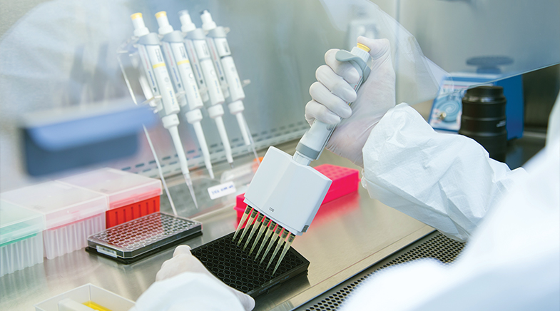
It begins with a 2-millimetre skin biopsy from behind the ear, when a device that works like a punching machine lifts off the surface layer of the skin. Skin cells are placed in plastic dishes with small wells, in sterile facilities, under special conditions that force them to change their identity. By the end of three months, the skin sample no longer resembles its former self. Its cells have become induced Pluripotent Stem Cells (iPSC), with the capacity to transform into almost any type of cell.
At the LV Prasad Eye Institute (LVPEI) in Hyderabad, through a process that lasts three more months, research scientist Indumathi Mariappan transforms the iPSCs into Retinal Pigment Epithelium (RPE) cells. The RPE cells first appear as black spots in the dish towards the end of a second three-month period. "We start with transparent iPSC cultures that become pitch black," says Mariappan. "That is the good thing about RPE cells. You can see that they are going in the right direction." The black cells are then selected and amplified into a group of RPE cells. Within six months of peeling the skin off the patient, the brown skin cells have transformed into black RPE cells.
Mariappan and her colleagues are developing RPE cells with the hope of creating treatments for retinal diseases, especially for those due to age-related degeneration. The epithelial cells are not directly involved in light sensing and image formation. This is done by rods and cones, which are photoreceptor cells in the retina. However, RPE cells nourish the photoreceptors, clear out the debris around them, and secrete growth factors that help the photoreceptors thrive. If the RPE cells start dying, photoreceptors start degenerating. And so does vision.
Cell therapies have become possible because of a discovery in 2007 by Japanese researchers Shinya Yamanaka and Kazutoshi Takahashi. The duo showed that by activating a set of four genes (later named as Yamanaka factors), any cell could be induced into an embryo-like state (bit.ly/Yamanaka-factors). These cells were called iPSCs and could be transformed into any type of cell. For example, you can take iPSCs, reprogram them into retinal cells, and put them into the eye – where they can replace malfunctioning or dead retinal cells.
Mariappan started working on developing RPE cell lines in 2008, right after she joined LVPEI. The Yamanaka factors had been reported just a year earlier, and she had been excited by the pursuit of iPSC-derived cell therapies for the eye. Nearly 15 years later, she has developed a robust method for making iPSC-derived RPEs. Animal trials will soon follow.
PAST ISSUES - Free to Read


Have a
story idea?
Tell us.
Do you have a recent research paper or an idea for a science/technology-themed article that you'd like to tell us about?
GET IN TOUCH


