Body of work
-
- from Shaastra :: vol 05 issue 04 :: Apr 2026
Regenerative humanised tissues could take the load off donor transplants and aid in patient recovery.
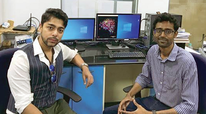
A meeting with an ophthalmologist set Pandorum Technologies on a new path. In 2010, aerospace engineer Arun Chandru and biophysicist Tuhin Bhowmick entered a contest, organised by the Department of Biotechnology (DBT) and the Association of Biotechnology Led Enterprises. The two had their sights on the ₹5 lakh prize money, with which they hoped to supplement their modest PhD stipends. They won the first prize in the Best Biotech Entrepreneurship category and, a year later, set up Pandorum, initially to produce 3D tissues of organs such as the liver for pharmaceutical companies to test their drugs on.
Those were the early years of the biotech boom. In 2012, the DBT established the Biotechnology Industry Research Assistance Council (BIRAC), and Pandorum was among the first to receive a grant of ₹50 lakh from the council. "We might have continued as a boutique company manufacturing organ models for testing had we not met the reputed ophthalmologist, Dr Virender Singh Sangwan," recounts Chandru. Sangwan, a cornea expert, encouraged the Pandorum team to focus on creating a transplantable tissue to address blindness. Fifteen years and several trials and failures later, Pandorum is ready to begin human trials with its two biomimetic liquid cornea products: Kuragenx and Kuragel.
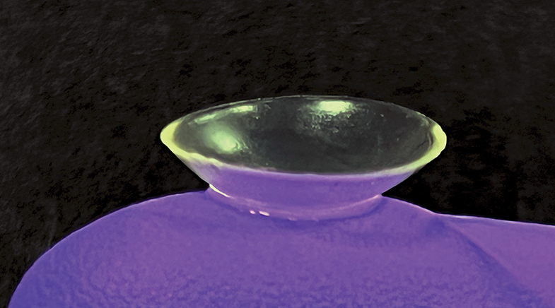
Pandorum created a hydrogel containing collagen and hyaluronic acid, with exosomes derived from corneal tissues.
Chandru recounts that before he met Sangwan, he knew little about eye transplants. "I thought the entire eyeball was transplanted," he says with a chuckle. Once they understood the biology, they realised they were in a field where technology was changing rapidly. They tried numerous methods, including 3D bioprinting, but ultimately achieved the best results by creating a hydrogel containing collagen and hyaluronic acid, with extracellular vesicles (exosomes) derived from corneal tissue. The gel, to be applied to the cavity formed when a damaged cornea is removed, is fixed with light. Being transparent, it mimics the corneal epithelium, while simultaneously providing a matrix for the eye's own corneal tissues to regenerate on.
In animal tests, the gel has shown a 90% regain of the original cornea's functionality. If commercially viable, the product could reduce dependence on cadaveric corneas and help resolve corneal blindness for many. The products have been granted 'Orphan drug designation' by the U.S. Food and Drug Administration (FDA). The designation helps developers get financial and tax incentives, regulatory support and research grants.
The first artificial cornea approved by the FDA was AlphaCor in 2002, but because it caused several complications, it fell out of use and is no longer manufactured. The global market leader is Boston KPro, but it, too, is recommended only when donor transplants are not advisable. It is a synthetic prosthesis.
REGENERATIVE PHILOSOPHY
The new thinking in bioengineering is to create prostheses that are not inert but aid the body's regenerative process. Developments in the past 15 years, with the easy availability of 3D bioprinting and other tools, and a deeper understanding of the role of structures such as exosomes, have helped advance the thinking. Bioprinting enables researchers to create highly complex architectures with precision. Exosomes are a game changer in tissue culture. They encapsulate all the properties of the parent cell, including the genetic information. However, they are easier to use because they are more stable and can be kept at room temperature; they can also be produced on a large scale. There is also a lower risk of exosomes turning tumorigenic or causing immune rejection.
Researchers also work on regenerating tissues, especially cartilage and bone, using a biodegradable polymer as a substrate for the tissue.
Across research institutes in India, scientists are working to create human-like tissues to replace damaged ones. While the low-hanging fruit is corneal tissue, which is avascular, and the bone, which has an easy-to-replicate architecture, scientists are also working to develop replacements for cardiac muscles, lung tissue, and even the food pipe. Such tissues are expected to replace much of the functionality of a damaged organ and are a step up from inert prostheses.
At the Indian Institute of Technology (IIT) Hyderabad, Falguni Pati, too, is developing a corneal tissue replacement. His team, in association with the LV Prasad Eye Institute and the Centre for Cellular and Molecular Biology, has developed a hydrogel which can be used as a bioink to print corneal tissue. The raw material is cadaver corneas, which are unfit for transplant. The matter is decellularised by removing the corneal cells. The remaining extracellular matrix containing exosomes is enriched with limbal stem cells, derived from the eye, which play an important role in rejuvenating corneal tissue. The resultant hydrogel is the ink with which the stroma and corneal epithelium are bioprinted ('The eye is a window to the body'; bit.ly/Shaastra-Eye). Pati's work has secured a grant from the Indian Council of Medical Research's 'First in the World Challenge' award for partial keratoplasty or cornea replacement. His team has conducted several rounds of successful animal tests.
In 2025, his lab began work on constructing a biomimetic oesophagus or food pipe. Cancers of the food pipe lead to patients having to live with compromised feeding after the malignant tissue is removed. The food pipe has two distinct tissues: a mucosal lining and smooth muscles. These muscles are arranged in an orthogonal fashion in layers so that they can constrict both radially and lengthwise, for peristaltic movements. "The level of complexity is higher; we also have to develop the vascular tree for such tissues," says Pati. His lab developed a technology for bioprinting muscles in different layers, and has filed for a patent, too, he adds. The research work is in association with AIG Hospitals, Hyderabad. They have cleared one level of animal testing.
CLOSE TO THE BONE
At IIT Kanpur, the Department of Biological Sciences and Bioengineering is abuzz with experiments to create a range of tissues, including regenerative bone cement, cardiac patches, and a newly launched project to regenerate nerve tissue. "We look at tissues that have the potential to regenerate but need support or scaffolds. Sometimes, if the possibility of regeneration is not there, then we see how to modulate it," says Ashok Kumar, Professor in the department. His focus is the skeletal system, and he addresses bone loss due to conditions such as trauma, tuberculosis, osteoporosis and necrosis.
A success story from the lab is a bi-phasic ceramic filler or cement made with nano-hydroxyapatite and calcium salts that naturally occur in bone. When injected into the injury site, the paste sets in 15 minutes. Apart from cementing the injury, it also acts as a scaffold for new bone cells to grow. "Defects as big as 2-3 cm long can be recovered," Kumar says. Over time, the scaffold degenerates, and the new tissue takes over. The prevailing method for treating such bone loss is autografting, or using bone from another part of the body, which entails two invasive surgeries.
IIT Kanpur has a tie-up with Conlis Global, a Canadian biotechnology company, to license and market the ceramic filler. "Such bone fillers could also be used as a carrier of medication for targeted therapy for conditions like tuberculosis,'' says Kumar, the Principal Scientific Adviser to the start-up.
Armed with a bunch of novel technologies for orthopaedics, Kaustav Chatterjee and Indranil Chakraborty from the Indian Institute of Science (IISc) in Bengaluru, started MedrinZ BIO, a deep medtech start-up. While the first product range for marketing will be implants and bone plates of titanium, priced much below those that are imported, the aim is to manufacture a line of bioengineered tissue products. ('Game, set, print'; bit.ly/Shaastra-Implants). "We look at orthopaedic solutions in various ways, from designing self-disintegrating rods, nuts and bolts to fix broken bones, to regenerating bone, cartilage and vascular networks within the body,'' says Chatterjee. His biomaterials and tissue engineering lab at the IISc is working on alloys of materials such as magnesium and zinc to create rods and plates for fracture repair. Unlike steel, these materials dissolve over time as the bone heals, eliminating the need for additional surgery to remove them. The researchers also work on regenerating tissues, especially cartilage and bone, using a biodegradable polymer, such as polylactic acid, as a substrate for tissue growth.
Biman Mandal's favourite substrate for bioengineering is silk protein. He prefers the non-mulberry wild silks of Assam, such as tussar, muga and eri. These silks have a unique tripeptide bond of arginine, glycine, and aspartate, called RGD (Arginylglycylaspartic acid) in extracellular matrix proteins, which allows cells to sit on them. "Other substrates like chitosan and cellulose need an attachment of collagen, or we have to do some chemistry on them to create RGD bonds. It is preferable to do minimum chemistry on a material to reduce issues of toxicity and rejection," says Mandal, Professor in the Biosciences and Bioengineering department of IIT Guwahati. While the lab works on bio-printing bone substrates, skin models, blood vessels and cardiac patches, Mandal is focusing on the spongy tissue in the hollow lining of long bones. The tissue has marrow that produces blood cells. "The hard structure of the bone is largely taken care of. We are now figuring out how to keep the marrow cells alive and functional. The environment for it is niche, and it is a challenge,'' he says.
Several research institutes around the world are working on creating functional marrow. Harvard University's Wyss Institute for Biologically Inspired Engineering is among the leaders, with studies on topics such as marrow-on-a-chip and injectable bone marrow scaffolds for transplanted haematopoietic stem cells. A study led by Queen Mary University of London details the creation of biomimetic bone marrow niches (bit.ly/marrow-niche). "The final frontier will be bioprinting a bone or a part of bone which is functional in every way, from providing skeletal support to manufacturing blood cells," says Mandal.
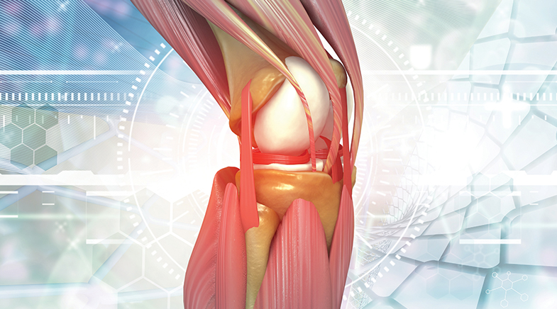
His team also works on regenerating cartilage and is looking to create a regenerative cartilage-bone interface that could be useful for patients with osteoarthritis. Using 3D bioprinting, his lab created a three-layered knee meniscal tissue that could be developed for transplantation in patients with meniscal damage, primarily due to sports injuries. An injectable gel version with a silk protein substrate can be used to repair partial tears on site, says Mandal. These technologies are at the animal-testing stage.
Exosomes are a game changer in tissue culture: easier to use, they are more stable and can be kept at room temperature.
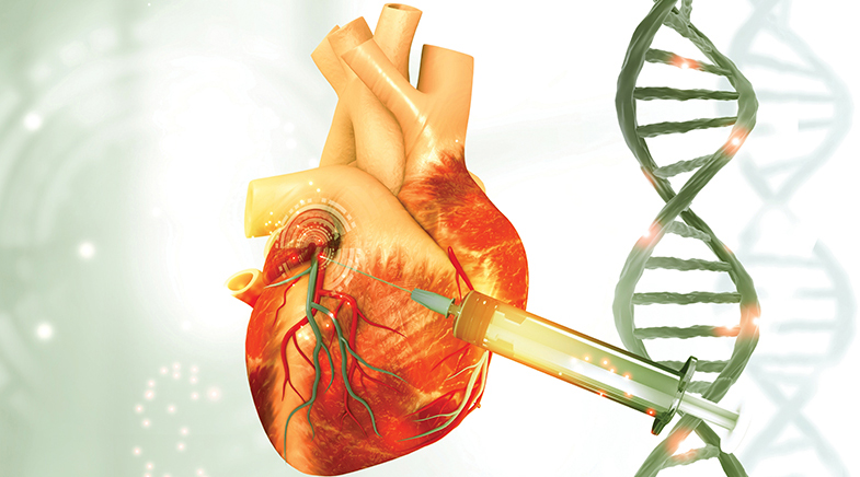
In a 2021 study conducted in collaboration with Harvard Medical School, the team created cardiac muscle patches capable of maintaining a rate of 75-78 beats per minute. These patches could restore the function of the heart ventricle with damaged muscles (bit.ly/beating-muscle). The heart is also a target tissue for the IIT Kanpur team. It is working on injectable gels to be administered directly at the site of fibrosis in the ventricle. The gel contains cardiac muscle cells in a collagen matrix.
Ashok Kumar's next target is developing scaffolds for nerve tissue regeneration. The idea is to provide some intervention for people with paralysis. "Our scaffolds have shown good results with animal tests," he says. Pati's lab has begun initial work with lung tissue. While creating entire organs for transplants may still be years away, researchers believe that within 5-7 years, there will be many commercially available tissues for treating patients. "The technology has reached a stage where it is racing," says Pati.
As an offshoot of the ongoing research, most labs are also developing humanised, biomimetic models to test drugs and cosmetics on ('Raising the bars' ). The need for such testing platforms is becoming imperative, with the U.S. National Institutes of Health announcing in 2025 that it would no longer fund projects based solely on animal testing models. India, too, revised its New Drugs and Clinical Trial Rules in 2023 to encourage non-animal-based platforms such as organs-on-chip, 3D biomimetic models and advanced computing methods.


Learning, unlearning
Every new tool — from a calculator to the GPS — has eroded human ability. Now, all eyes are on AI.

Ideas with a vision
Easing ECG tests, cotton farming and lab lessons.

To the Moon and back
A ready reckoner for Artemis II's Moon Mission.

Have a
story idea?
Tell us.
Do you have a recent research paper or an idea for a science/technology-themed article that you'd like to tell us about?
GET IN TOUCH